Invasive animals are non-native species that survive in a variety of environmental contexts, and whose range expansions cause economic or environmental harm. Understanding how certain species become invasive can help mitigate their spread and colonization.
Many invasive vertebrates, especially fishes, have specific adaptations that enable them to tolerate extreme environmental conditions or move into new territories. One adaptation that may facilitate invasive behavior is body shape. An eel-like body plan has evolved multiple times in fishes, underscoring its evolutionary advantages. Researchers know that highly elongate fishes are able to move forward and backward more readily and traverse the water-land interface to move on land for periods of time.
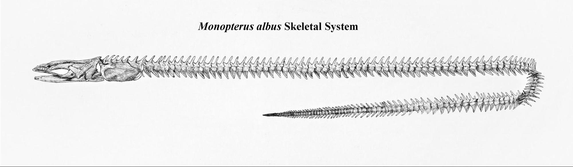
 For my project, I examined the internal anatomy of the Swamp Eel, Monopterus albus, with dissections and scientific illustration in order to determine how the internal anatomy differed from other elongate fish and how this anatomy may facilitate adaptation to new environments. The Swamp Eel is not related to “true eels” such as American Eels, Moray Eels, or Conger Eels but has independently evolved an eel-like body plan. The Swamp Eel is native to Southeast Asia and has invaded the Northeastern and Southeastern United States. I created three illustrations that depict the entirety of the internal anatomy and the skeletal system of this invasive fish. Through my dissections and illustrations, I compared the internal anatomy of the swamp eel with other unrelated elongate fishes.
Dr. Andrea Ward from Adelphi University sent us six Swamp Eel specimens ranging from 629 - 766 mm in total length. After exposing the body cavity, I recorded organ lengths and placed pins through the vertebral column at the anterior and posterior position of each organ to mark their vertebral spans, which is a proxy for organ size. I took photos to document my dissection process and to use as a reference for my illustrations. Dissected specimens were dried in the fume hood. Once dried, I counted the total number of both body and tail vertebrae and indicated the position of the pins so I could determine the vertebral span of each organ. Once all data were collected, the pins were removed and the specimen was placed in a dermestid beetle colony. Dermestid beetles will eat all of the soft tissues, leaving only a skeleton behind as an osteological specimen. The osteological specimens are archived in the Mehta Lab.
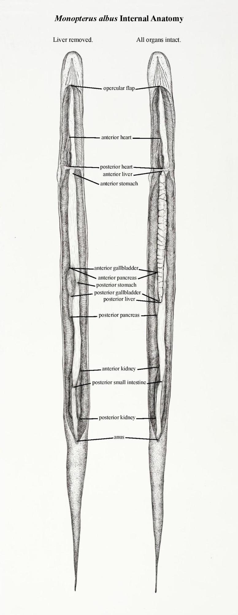
The Swamp Eel is a fascinating creature, with an array of interesting characteristics. The Swamp Eel lacks scales, is greenish-brown in color, and has countershading, with a lighter underside. The eyes are small and are positioned near the front of the head. Swamp Eels differ from most other fish because they have a single opercular opening on the ventral side of their body, as opposed to most fish which have paired openings positioned on each side of their head. The Swamp Eel uses its highly vascularized mouth cavity to breathe and can survive on land for an extended period of time. It lacks any specialized air sacks so the buccal cavity, pharynx, and anterior esophagus are highly vascularized to facilitate gas exchange. The Swamp Eel has an average of 101 body vertebrae and 56 tail vertebrae, which is unusual as most eel-like fishes have many more tail vertebrae. Most of the organ systems of the Swamp Eel (heart, liver, gastrointestinal tract), are significantly larger than other elongate fishes even when correcting for their large body size. Larger organs need more oxygen. Presumably the Swamp Eel’s ability to breathe air through its mouth cavity may help it oxygenate its large organs in anoxic aquatic conditions.
I completed two illustrations documenting the viscera of Swamp Eel, one containing all of the organs and one with the liver removed to expose the organs underneath. Through this project, I gained a deeper understanding of the anatomy and physiology of Monopterus albus and have documented what I have learned through these illustrations. Whole anatomical diagrams of this invasive species are lacking in the literature. My illustrations will be useful to anyone wanting to gain a better understanding of this elongate invasive fish or using a comparative anatomical approach. I would like to thank the Norris Center for funding my work.